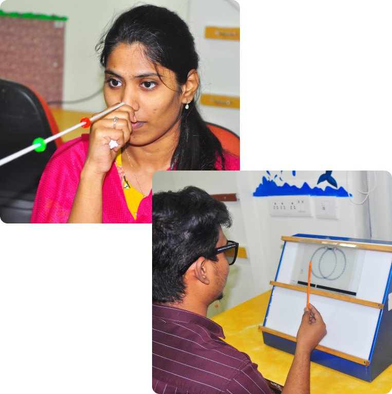
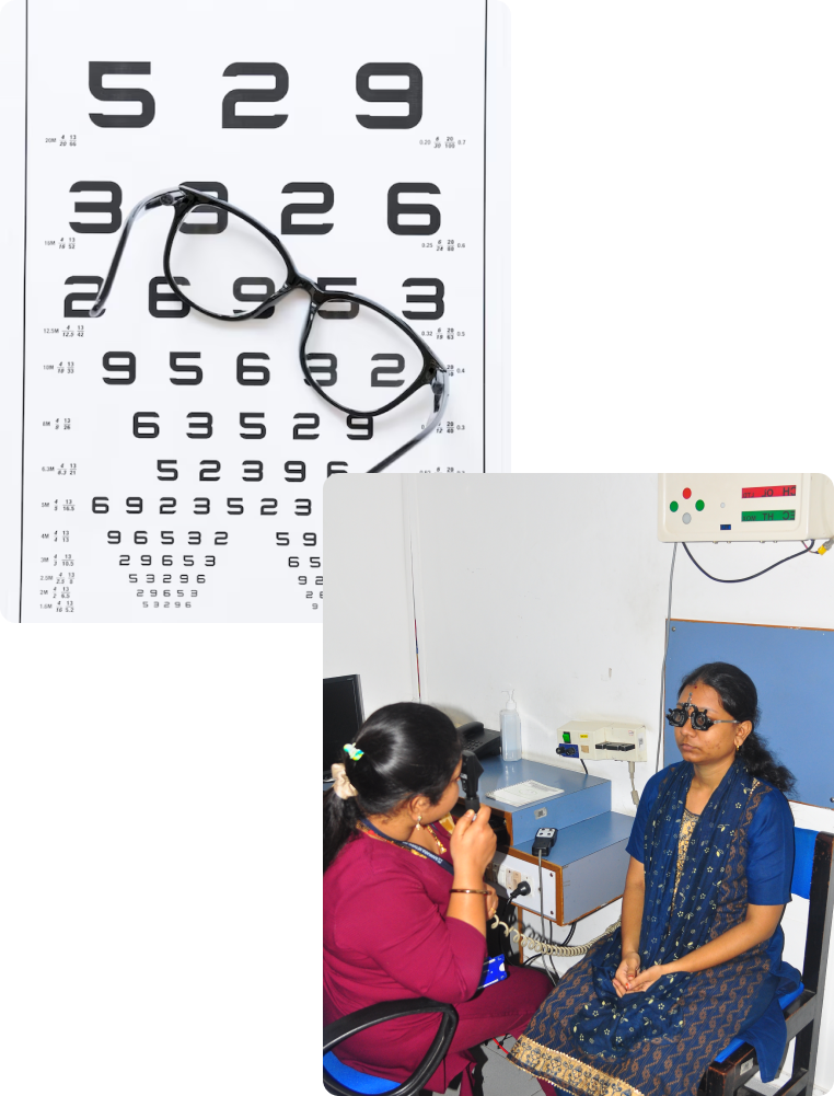
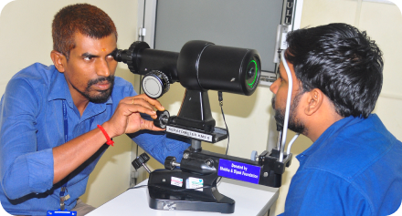
We offer both out-patient services and in-patient services at our hospitals. We have a dedicated team of Specialist Ophthalmologists, nursing staff, administrative staff and support staff...
We have a full fledged drug store in the hospital premises, where our patients can buy the drugs prescribed by the consultants without having to go outside....
Our hygienic Canteen within the hospital ensures healthy food to the patients and to the attendants. It provides South Indian dishes with additional touch of north, east and west Indian food items...
This section will help you in better understanding the services and facilities available at Sankara Nethralaya and in understanding medical terminology that your doctor would be using at the time of examination and treatment of your eye, and help to raise your Eye Q
Vision can be defective to a variable degree. It may be easy to detect gross decrease in vision but it may be more difficult to detect subtle degree of loss of vision. It is very easy to miss gross loss of vision in one eye when the other eye is healthy unless one consciously tests each eye separately. It is a good practice to test each eye separately at regular intervals using any fine reading material such as newspaper.
Normally the image formed by the two eyes is coordinated into a single image by the brain. Two distinct images are seen once this coordination is disturbed due to various diseases involving the muscles of the eye and the nerves that control the same. Multiple images often are an early symptom of cataract.
Visible redness of the one or both eyes is a common symptom pertaining to several varieties of diseases. One should not mistake every red eye as having viral conjunctivitis (So called Madras eye). Hence do not self medicate and delay seeking medical advice if you have a red eye. It could be something serious.
A common symptom of infection in the eye is stickiness of the eyelids due to discharge. This infection could be purely external or could be more serious. Persistent stickiness of the eye lashes needs early evaluation.
Watering could be the result of mal alignment of the eyelids or eyelashes or a blockade of tear ducts that normally drain the tear fluid into the nose. Presence of tearing in newborn babies can indicate lack of patency of the tear ducts and may need attention.
Normally the center of the eye gives a black reflex due to the pupil. A white reflex can be due to opacification of the normally transparent cornea, the lens (cataract) or due to an abnormal growth of tissue behind the lens. A white reflex in a child can potentially be dangerous and should not be ignored.
Abnormal look of the eye could be due to prominence of the eye, or could be the result of defects involving the eyelids. Prominence of the eye could be due to large eyeballs or due to protrusion of normal sized eye by abnormal growth behind the eye. Any change in appearance of the eye should be investigated. Previous photographs could be useful in comparing especially when one is not certain about the time of onset of the abnormal look of the eye.
Drooping of the upper eyelid could be present at birth or could occur later. If the defect has occurred later in life one should note the frequency of the occurrence and in what part of the day it is more prominent. These observations can help the doctor make important decisions.
Squinting indicates the misalignment of the eyes. In children, this can potentially lead to reduction of vision in the squinting eye due to disuse (lazy eye). When in doubt, taking photographs with flash can help identify the squint in the photographs. This is especially useful to the doctor, in case of children who refuse to cooperate with the doctor for adequate examination.
Vision testing involves making a person read standard sized letters at a specified distance. The Optometrist record the vision as a fraction e.g. 6/6 etc. The top number denotes the distance (in feet) at which the patient has been able to read the particular sized letter while the bottom number indicates the distance at which a normal person is expected to read the same letter. Near vision is tested separately in good illumination using special test charts held at normal reading distance. The testing is done with each eye separately. The Optometrist often test the vision using a pinhole. This gives an estimate of improvement possible with glasses. The patient in place of glasses cannot use the pinhole.
This is an important test that is done by the Optometrist. The eye is like a camera. The light rays are focused on to the light sensitive film in the back of the eye called the retina. This focusing is made possible by the cornea (a clear watch glass like structure in the front of the eye) and by the lens in the eye (similar to the lens of a camera). Refraction is done usually in the normal state. On occasion (especially in children) it may have to be done using special eye drops (cycloplegics). In some circumstance one may have to retest the power of the required glasses 2-3 days after the testing with the use of drops. Refraction involves two parts. The first part is objective where in the Optometrist estimates the power needed by using a test called retinoscopy. This test can also be done with a machine called the auto-refractometer (so called computer testing). However, one still needs to do the all-important subjective testing (i.e. testing the response of the patient with different powered glasses) before prescribing the glasses.
This test is done in selected group of patients depending upon their symptoms. The test involves looking at a chart that has a grid drawn with a central dot. The test is done using the near vision glasses (if one is using the same). The test permits the evaluation of function of the central 20 degrees of the retina. The patient is asked to look at the central dot and tell whether
All the corners of the chart are seen All the lines are seen straight and not crooked There are any areas of dark patches where the lines are not seen. Whether the central part of the chart or the peripheral part of the chart is clear. The Amsler's chart is very useful as a home monitoring device. If any defect is noted, immediate ophthalmologic examination is warranted.
One of the most common procedures that is done in an eye specialist's office is dilatation. The pupils of the eye constrict or dilate depending upon the light that thrown at the eye. For examining the back part of the eye (fundus), the doctor uses an instrument called ophthalmoscope. To get a good view of the back of the eye, one needs to dilate the pupils. This permits more light to enter the eye and gives a better image of the fundus. To keep the pupils dilated despite the intense light, one needs to dilate the pupils. There are various types of dilating drops available. The faster acting ones may dilate the pupil in 15-20 minutes time. Other variety of drops may take up to 30-45 minutes for good dilatation. The effect of dilatation usually lasts up to 6 hours. Some of them may retain the effect for 24 hours. Usually, the drops used for routine eye examination do not have long lasting effect. A patient is expected to have glare in the sun light while still under the effect of the drops. Hence driving may become difficult. If you had similar dilatation in the past and have been noted to be allergic to any one of them, please inform the same to your doctor.
Slit lamp is an instrument that has an in-built microscope and a bright illumination system. The special arrangement of the light and the microscope allows the Ophthalmologist and Optometrist to view the eye in great detail under high magnification. The front part of the eye is examined without any other aids while the back part of the eye (fundus) is examined with help of special lenses held in front of the eyeball.
Tonometry involves the check of the pressure of the eye. Normal pressure is a range and not a finite number. Raised pressure in the eye can be harmful to the nerve connecting the eye with the brain (optic nerve). The pressure is normally checked in one of the three ways.
Here a metallic device is placed on the eyeball and the deflections of the needle on the scale are used as a guide to measure the eye pressure. At the best, this modality of testing can be used only as screening device. This is not very accurate.
In this a small prism mounted on the slit lamp is used to contact the eyeball and measure the pressure. This modality of testing is more accurate and is the standard today.
The Perkins tonometer was devised as a portable handheld applanation tonometer, for use in children, patients unable to cooperate for slit lamp examination, and anesthetized and bedridden patients. PAT is currently in widespread use in the primary care setting.
This is an electronic device that is very accurate and useful in specialized situations.
A drop of anesthetic is placed on the eye before these tests are carried out. The test is totally painless.
ICare 200 is an updated version of rebound tonometer for measuring intra-ocular pressure. It is very useful is measuring IOP for children, bedridden patients and non-cooperative patients for GAT. It can measure IOP in both supine and sitting position and reliability is also almost equal to GAT.
Gonioscopy is the test in which the angle of the eye is examined. A fine balance between the inflows of fluid maintains the eye pressure and it's outflow. The outflow is through the angle of the eye. Studying this angle gives a lot of insight into the cause of a condition called glaucoma. This test is done with the help of the slit lamp and a special lens called gonioscope. The eye is anesthetized by placing a few drops of anaesthesia to facilitate placement of the lens. This test is totally painless.
This is a very important step in the total examination of the eyes. The visible portion of the eye is easily examined by the slit lamp examination. The back portion of the eye can only be examined by using the ophthalmoscope. This step usually needs dilatation of the pupils. This test involves throwing bright light into the eye and examining the image of the back of the eye using special lenses. For Indirect opththalmoscopy, the patient has to be in the reclining position for proper examination. Sometimes the slit lamp may be used for detailed evaluation of the areas of the back of the eye such as macula, optic disc etc.
Potential acuity meter testing enables one to have an idea about the possible visual recovery following cataract surgery. In this testing the doctor projects a chart of letters or numbers into the back of the eye through the gaps in the cataract that enables the patient to read the letters. Depending upon the number of lines that one could read, the potential for recovery of vision is estimated. One should realize that this is only an approximate estimate and very often the true recovery of vision is greater that the estimate.
The glare testing permits one to assess the deterioration in vision that occurs with glare. Cataract can produce significant scattering of light. Hence people with early cataract may have good vision under ideal conditions of testing but the vision may deteriorate rapidly under conditions of glare. This testing enables one to decide on the need or otherwise for cataract surgery.
This is a very important test that enables one to calculate the desired power of intraocular lens during the cataract surgery. This artificial lens implanted in the same location as the natural lens permits one to have good vision without needing to use the thick glasses or contact lenses after cataract surgery. The test involves use of ultrasound to measure the length of the eyeball and this information along with keratometry (the measurement of the curvature of the cornea) is used to calculate the IOL power by a complicated formula.
Schirmer's test is a measure of the tear secreting capacity of the eye. Deficiency in tear secretion can lead to a chronic condition called dry eye. The test involves placement of a special filter paper strip across the lower eyelid margin and measuring the length of the strip that is wetted by the tears over a one-minute period.
Keratometry involves the measurement of the corneal curvature in two meridians. The cornea is the front portion of the eye that is clear like a watch glass. The curvature of the cornea helps it to focus the light partly. Measurement of the corneal curvature is needed for fitting proper contact lens. It is also needed for the calculation of the IOL power before cataract surgery.
A diagnostic tool, unveils the complex landscape of the cornea, offering a detailed map of its surface. Through advanced computer analysis of multiple points on the corneal surface, this technique utilizes the study of reflected images to provide comprehensive insights into the cornea's structure.Corneal topography plays a pivotal role in cases where corneal scars or sutures are present. By analyzing the surface map, ophthalmologist gain essential insights into the impact of these conditions on the cornea.This knowledge empowers them to make informed decisions regarding further treatment strategies or the removal of sutures.
It involves the scanning of both the front and back surfaces of the cornea, capturing cross-sectional images from all angles through a rotating camera.This comprehensive approach provides a 360-degree view.One of Pentacam's standout features is its ability to diagnose corneal conditions, such as Keratoconus, even in its early stages.By utilizing advanced software, Pentacam assists in the early detection of abnormalities, allowing for timely intervention and effective management.Additionally, it serves as a crucial tool for follow-up assessments in the ongoing treatment of corneal diseases.
Pachymetry is the study of the thickness of the cornea. The accurate measurement of the thickness is made possible by using ultrasound or optical means. Measurement of the thickness is important in the diagnosis and management of certain corneal conditions such as keratoconus, corneal endothelial dystrophy etc.
Specular microscopy is a test that enables the evaluation of the back most layer of the cornea called the endothelium. The health of this layer is important in maintaining the clarity of the cornea. With age, injury, surgery and in some diseases this layer may have reduced number of cells and become abnormal. Study of this layer is done by counting the number of cells per square millimeter as well as study the type of the cells. This study is important in planning certain surgeries.
Schirmer's test is a measure of the tear secreting capacity of the eye. Deficiency in tear secretion can lead to a chronic condition called dry eye. The test involves placement of a special filter paper strip across the lower eyelid margin and measuring the length of the strip that is wetted by the tears over a one-minute period.
Field of vision describes the side vision when one is looking straight ahead. The testing of the extent of the side vision is important in the diagnosis and follow up of several disease including glaucoma, and diseases relating the eye with the brain (neuroopththalmology). The test is usually done on computerized machines (Humphrey field analyzer). The machine is programmed to test several points, sometimes repeatedly with varying illumination. The test may take time depending upon the defect in a given patient. The computer has inbuilt software that enables comparison of the field charting on repeat testing of the same patient.
Optic disc is the only part of the optic nerve visible to the eye doctor in the back of the eye. The appearance of the disc gives valuable information to diagnose and treat conditions such as glaucoma. It is important to be able to compare the appearance of the disc over a period time in cases of chronic glaucoma. This is made possible by several techniques- one of which is the photography of the disc using the fundus camera.
This recent innovation allows measurement of the thickness of the nerve fibre layer, which is the part of the retina that is first affected in the disease of glaucoma. The nerve fibril layer defect is detected long before any defect is noted in the function of the eye including the field examination. This test may help in the early detection of significant damage caused by glaucoma and help in the follow up of these patients. The test involves the use of scanning laser that passes through the nerve fibre layer and in the process undergoes a process called retardation. By measuring the extent of retardation the machine calculates the thickness of the nerve fibre layer.
This is an advanced technology in ultrasonography, which permits high-resolution pictures of the front of the eye. The technology enables the measurement of the angle of the eye, which is otherwise not accessible for measurement. The angle of the eye is the path through which the fluid in the eye finds access outside. The angle can become closed in certain individuals. This propensity to closure of the angle can be more adequately predicted using this advanced testing. Following injury to the eye, sometimes abnormal communications develop leading to excess drainage of fluid and resulting soft eyes. These abnormal sites can be best identified by UBM.
HRT (Heidelberg retinal tomography) is an imaging technique developed with the objective to reliably measure and follow structural changes in optic nerve head or the thickness of RNFL in glaucoma patients. HRT is a one such imaging modality, uses a confocal scanning laser to image serial sections of optic nerve to create a map which is useful in glaucoma patients.
These two tests enable the measurement of misalignment between the two eyes. This type of problem leads to a condition of double vision in a patient. The extent of the double vision and the direction in which it is maximal can be charted by using these two tests. The tests are done using red and green goggles where in one colored glass is placed in front of one eye and the other in front of the other.
Certain disease of the retina and optic nerve leave behind subtle defects of sensitivity. A patient is very symptomatic of these deficiencies but the commonly performed tests like the vision testing do not reveal the true extent of the defect. Measurement of contrast sensitivity enables one to understand these subtle defects in the visual function. This test involves identification of patterns of gray on gray background.
Color vision is an important component of human vision. Defects in this can be by birth or due to any acquired diseases. The testing is done using one of the two methods.
Ishihara's charts- In this, many charts are presented and the patient is asked to identify the letters or numbers in the chart. Farnsworth- Munsell 100 hue test- In this the patient is asked to arrange several caps of different hues in their order. The test is done in good illumination.
In this test bright light or patterns of dark and light bands are projected on to the eye. The electrical potentials that are generated in the brain as a result of the light or pattern are recorded. This gives valuable information regarding the functional intactness of the optic nerves and the optic pathways that normally conduct these impulses to the brain
CT scanning is a computerized system where in x- rays are used to construct images of thin slices of tissues allowing detailed evaluation of the tissues under consideration. By manipulating the soft ware, the image quality and detail can be enhanced. Injecting some drugs called contrast agents can get additional information. CT scanning is very useful in the evaluation of diseases of the orbit (bony cage in which the eye is located) as well as some diseases of the eye itself. Injury related problems - especially presence of foreign bodies is easily picked up and located on CT scanning.
MRI scanning is a different technology and looks at the tissues in a different perspective. Sometimes both CT and MRI scanning may be needed to understand some diseases. MRI is especially useful in diseases of brain that may affect the eye. By using some specialized soft ware, one can even image the blood vessels of the brain without injecting any drug ( MR Angiography).
Fundus photography permits documentation of the structures of the eye. This documentation may be important to compare with other investigations such as fluorescein angiography as well as for follow up. Fundus photography of the optic disc is important in the management of glaucoma.
This is an important test to evaluate a variety of retinal disease such as diabetic retinopathy. This is one of the commonest tests performed for retinal diseases. The test involves injecting a dye called sodium fluorescein into the blood stream and taking photographs of the retina using special filters. The test is important to stage the disease as well as to guide treatment with laser photocoagulation. Present generation digital cameras permit manipulation of the pictures and for instant viewing without need for development of the film etc.
Indocyanine angiography is similar to the fluorescein angiography but involves injection of a different dye called Indocyanine green. The test utilizes a special infrared sensitive camera to capture the images digitally. Very often indocyanine and fluorescein angiography are combined in a given patient to give maximum information. Indocyanine angiography gives more information regarding the choroidal vessels compared to fluorescein angiography that gives more information regarding the retinal blood vessels.
These two tests are done to evaluate the function of the retina. Light is projected onto the retina and electrical potentials that occur normally in the eye are recorded using special electrodes placed near and on the eye. Certain retinal degenerative diseases are diagnosed only on testing with electroretinography. Specialized computer soft ware is needed to analyze the data.
There are certain diseases that may lead to permanent partial loss of vision. These patients can be sometimes helped to some extent by using special aids called low vision aids. There are a variety of these available and most of them are fine tuned for a specific function. Most of them have been made to enable reading fine print. It is important that the patient should be motivated to use them. They are used at a closer range than normal working distance and hence one needs to get used to the same. Computers and closed circuit television are also useful as low vision aids. One has to test different varieties before choosing what is appropriate for them.
Ultrasonography is test that permits the evaluation of the back of the eye in case of opaque media. In a normal eye one is able to see the back of the eye using instruments such as indirect ophthalmoscope. In conditions of disease and injury the cornea, the lens or the vitreous cavity can become opaque and prevent this visualization. In these circumstances, ultrasound can be used to scan the eye and get useful information about the tissues lying behind the opaque media. This information is needed not only for proper diagnosis but also to plan the surgery where indicated.
An important part of the evaluation of a patient with squint is the cover test and prism tests. These tests are conducted in the office of the eye doctor itself. Using these tests the eye doctor is able to classify the type of the squint and to grade the severity of the same. This information is needed to plan the treatment including surgery where needed. With a torch light and a set of loose prisms the eye doctor is able to evaluate squint to a great degree.
Orthoptics is the study of how effectively the two eyes function together (binocular vision). This testing is done on many instruments including the synaptophore.. Special instruments are used to measure the near point of convergence and near point of accommodation. These points give a guide as to how difficult it is for a person to view near objects. Many people with eyestrain on performing near work may be helped by exercises after the orthoptic evaluation.