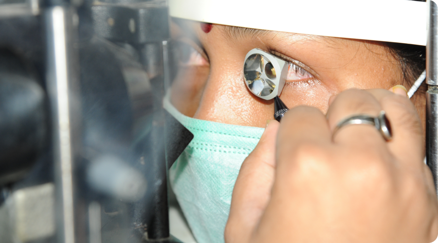
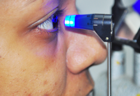
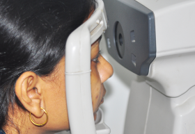
Investigative facilities
Our investigative facilities include an array of sophisticated equipment
- Ocular Surface
- Cornea
- Cataract
- Uvea
- Glaucoma
- Vitreo- Retina
- Neurological conditions
Ocular Surface
- Meibography
- Lipiview
- Anterior Segment – OCT
- Pentacam
- HRT3 Rostock Cornea Module
Cornea
- Corneal Topography
- Anterior Segment – OCT
- Pentacam
- HRT3 Rostock Cornea Module
- Specular Microscopy
- Aberrometry
- Pachymetry
Cataract
- A- Scan
- B- Scan
- IOL Master
Uvea
- Anterior Segment – OCT
- FUltrasonography
- Ultrasound Biomicroscopy
- Fundus Photography
- Posterior Segment OCT
- Swept Source OCT – Anterior and Posterior segment
- OCT - Angiography
Glaucoma
- Tonometry
- Gonioscopy
- Pachymetry
- Anterior Segment – OCT
- Pentacam
- Specular Microscopy
- Ultrasonography
- Ultrasound Biomicroscopy
- Visual Fields - Humphrey Visual field analyzer
- Optic Nerve head imaging – Photography and OCT
- Swept Source OCT – Anterior and Posterior segment
- OCT - Angiography
Vitreo-Retina
- Ultrasonography
- Visual Fields - Humphrey Visual field analyzer
- Fundus Photography
- Posterior Segment OCT
- Swept Source OCT
- OCT – Angiography
Visual Psychophysics
- Full field Electroretinography (ERG)
- Multifocal Electroretinography (mfERG)
- Electrooculogram (EOG)
- Pattern Electroretinogram (PERG)
- Visually Evoked Responses (VEP)
- Multifocal Visually Evoked Responses (mfVEP)
Neurological disorders
Functional Tests
- Contrast
- Colour Vision – FM 100
- Potential Acuity Meter
Electrodiagnostic Tests
Visual Fields - Humphrey Visual field analyzer
Optic Nerve head imaging – Photography and OCT
Lasers available
- ND YAG laser (Zeiss)
- Argon Laser (Zeiss)
- Diode Laser(Iridex)
- Lumenis SLT/YAG integrated combo Laser (Selecta Duet)Endoscopic Cyclophotocoaglation