| Back to Patient Care |
| Investigative |
|
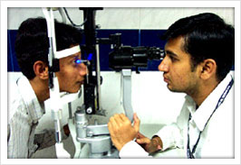
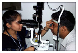
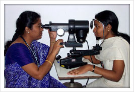
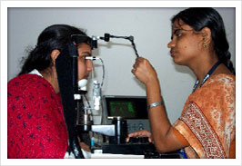
| Our investigative facilities include an array of sophisticated equipment |
|
| Cornea |
| Glaucoma |
| Vitreo retinal service |
| Neuro Ophthalmology evaluation |
| Imaging Techniques |
| Biometry – DBR (A Scan) |
| Electro-diagnostics |
| The Visual Fields (Perimetry) |
|
| Cornea |
|
|
Corneal Topography |
|
Specular Microscopy |
|
Aberrometry |
|
Pachymetry |
|
|
| Glaucoma Diagnosis |
|
Glaucoma is a disease where there is a raise in the intra-ocular pressure, optic disc changes and an associated visual field loss. |
| Intra - ocular pressure measurement |
|
| |
|
| |
| Applanation Tonometry |
| Non Contact Tonometry |
| Pascal DCT |
| Tonopen |
| Perkins tonometer |
| Proview |
| Reichert Ocular response analyzer |
| Rebound tonometer |
|
|
| |
| Anterior segment Imaging ; Visante AS-OCT (Zeiss) |
| Optic nerve evaluation by |
|
|
| Digital Fundus photography (Zeiss) |
| GDx VCC nerve analyzer (Zeiss) |
| HD Optical Coherence Tomography (Zeiss) |
| Heidelberg retinal tomogram |
| Ultrasound Biomicroscopy |
| Swept source OCT |
| Visual field testing by |
|
| |
|
| |
| |
| Humphrey Visual field analyzer |
| Frequency Doubling Perimetry |
| Blue on yellow perimetry |
| Differential light sensitivity |
| |
|
|
| Based on the above tests the type of glaucoma is diagnosed. |
| Lasers available: |
| ND YAG laser (Zeiss) |
| Argon Laser (Zeiss) |
| Diode Laser(Iridex) |
| Lumenis SLT/YAG integrated combo Laser (Selecta Duet) |
| Endoscopic Cyclophotocoaglation |
Remember
Glaucoma is the second most blinding disease in the World. It is known as the "Silent thief of sight" as most of the types of glaucoma are asymptomatic |
|
| Vitreo retinal service |
|
|
Ultrasonography |
|
Ultrasound Biomicroscopy |
|
Fundus fluorescein angiography |
|
Ophthalmic photography |
|
Electro diagnostic tests |
|
Optical coherence Tomography (OCT) |
|
|
| Neuro Ophthalmology evaluation |
|
|
Visual Psychophysics |
|
Contrast Sensitivity |
|
Color vision – FM 100 hue test |
|
Potential acuity meter test |
|
|
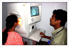
| Imaging Techniques |
|
|
Slit Lamp Photography |
|
Fundus Photography |
|
Fundus Angiography |
|
|
| Digital Biometry |
|
Purpose: |
| To determine the power of the intraocular lens (IOL) to be implanted in patients undergoing cataract surgery. |
| It involves measurement of the axial length (the distance between the anterior and posterior part of the eye) and the corneal curvature (curvature of the black portion of the eye ball) of the eye. |
| Test is done in both the eyes. |
|
| Keratometry: |
| Generally corneal curvature is measured first using an instrument called keratometer. |
| The patient needs to fixate steadily centrally as instructed by the examiner and the readings are obtained. he eye. |
| This is a non contact procedure. |
| A Scan: |
| Axial length is measured using ultrasound A scan |
| A drop of local anesthesia is instilled on to the eye |
| The patient is asked to fixate centrally |
| Examiner gently places a probe on the eye |
| Ultrasound waves are sent to the eye through the probe |
| The axial length readings are displayed on to a screen. |
| The examiner selects the appropriate reading |
| IOL power calculations are then done accordingly |
| Axial length measurements are also done in few other cases like |
| High myopia (high minus powered glasses) |
| Anisometropia (difference in spectacle power between both the eyes) |
| Nanopthalmos |
|
|
| Electro-diagnostics |
|
1. Full field Electroretinography (ERG): Test which measures the electrical responses of various cell types in the retina, including the photoreceptors (rods and cones), inner retinal cells (bipolar and amacrine cells). Used to diagnose various retinal degenerations. |
| |
Retinitis pigmentosa and their variants |
| |
X-linked juvenile retinoschisis |
| |
Heredo-macular degenerations |
| |
Retinal Vascular occlusions |
| |
Intraocular Foreign Body |
|
| 2. Multifocal Electroretinography (mfERG): Multifocal electroretinography (mfERG) is a valuable technique in assessing macular function in retinal disease objectively. It is used to record separate responses for different retinal locations. It is also used in the detection of |
| |
Macular dystrophies |
| |
Macular hole |
| |
X-linked retinoschisis |
| |
Drug toxicity |
| |
Multifocal choroditis |
| |
White-dot syndrome |
|
| 3. Electrooculogram (EOG): This is used to assess the function of the outer retina and Retinal Pigment Epithelium (RPE). EOG is used to confirm |
| |
Best disease |
| |
Suspected drug toxicities |
|
| 4. Pattern Electroretinogram (PERG): This test provides information about central macular and retinal ganglion cell layer. It is also used to differentiate vision loss due to retinal or optic nerve diseases. PERG is used in evaluating |
| |
Glaucoma and ocular hypertension, |
| |
Optic neuritis other optic neuropathies, |
| |
Maculopathies |
|
| 5. Visually Evoked Responses (VEP): The VEP is a test to detect problems with the optic nerve and lesions in the anterior part of our visual pathway. VEP tests are used to evaluate |
| |
Optic neuritis, |
| |
Compressive Optic neuropathy, |
| |
Toxic amblyopia |
| |
Cortical Blindness |
| |
Demyelenating diseases such as multiple sclerosis. |
| |
Unexplained visual loss |
|
| 6. Multifocal Visually Evoked Responses (mfVEP): This test allows for topographical assessment of visual field. In this test multiple individual VEP responses are recorded simultaneously from 60 or so regions of the central 20 to 25° radius of the visual field. This is also known as objective visual field perimetry. Indications are |
| |
Diagnosing and Following of Optic Neuritis/Multiple Sclerosis |
| |
Unexplained visual loss |
| |
Detecting and following of Glaucomatous damage |
| |
Confirming unreliable or questionable fields |
|
|
| The Visual Field |
|
What is Visual Field?
Visual field is the degree of side or peripheral vision a person can perceive when looking straight at an object.
How is the Visual field of a patient measured?
Perimeter is the instrument used to measure the visual fields of a patient. There are various types of perimeters to quantify the visual fields.
What instrument do we use at our hospital?
Perimetry is performed in Sankara Nethralaya using the AUTOMATED HUMPHREY FIELD ANALYZER (HFA II and HFA i-SERIES)
Why is Visual field testing important?
The purpose of visual field testing or perimetry is to provide information critical to: |
| Diagnosing ocular diseases, especially Glaucoma |
Evaluating neurological diseases
|
| Monitoring the progress of ocular and neurological diseases. |
| How is the visual fields testing done? |
| Mostly done for one eye at a time followed by the other eye. |
| Sometimes visual field testing is done for both the eyes simultaneously which is known as Binocular Visual field testing. |
| A perimetrist will assist the patient by clearly giving the instruction on how to perform the testing. |
| Patient’s understanding of the test and cooperation is very important to get a reliable report which in turn will assist the Ophthalmologist in the diagnosis and management of the disease. |
| Hence visual field plays a major role in early detection and treatment of the disease and also in evaluating the efficacy of the therapy used to control the disease process. |
| Normal fields |
| Inferior arcuate defect in glaucoma case |
| Superior arcuate defect in glaucoma case |
| Biarcuate defect in glaucoma case |
| Full Field 120 in a retinal case depicting central scotoma |
| Neuro case of Left Homonymous Hemianopia. |
| Neuro-OS |
| Neuro-OD |
|