| Molecular Microbiology |
Molecular Microbiology Tests Based On PCR Offered At ABSN Kolkota Molecular Microbiology Laboratory |
| Polymerase Chain Reaction Tests (PCR) |
| |
 |
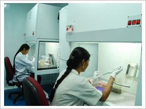
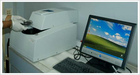
| Amplification Room |
|
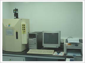 |
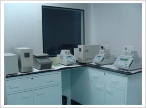
| Post PCR Gel Documentation Room |
|
|
| |
|
| |
| Nested PCR |
| Eubacterial genome |
| Panfungal genome |
| Propionibacterium acnes |
| Propionibacterium acnes |
| Cytomegalovirus(CMV) |
| Herpes Simplex Virus (HSV) type 1 & 2 |
| Toxoplasma gondii (T. gondii) |
| Chlamydia trachomatis |
| Chlamydia pneumoniae |
| RT PCR Rubella Virus |
| Adenovirus |
| Varicella Zoster Virus (VZV) |
| HLA-B27 Typing |
| REAL TIME PCR TESTS |
 |
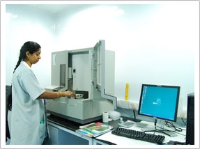
Real Time PCR Machine
(Corbett Rotor Gene) |
|
| Real Time PCR For |
| Mycobacterium tuberculosis |
| Human Immunodeficiency Virus |
| Chikungunya Virus |
| Human Cytomegalovirus (CMV) |
| Hepatitis B Virus (HBV) |
| Hepatitis B Virus (HBV) |
| Herpes Simplex Virus (HSV) |
| RAPID NUCLEIC ACLD – BASED MOLECULAR DIAGNOSTIC PROCEDURES FOR RARE & DIFFICULT TO DIAGNOSE INFECTIOUS DISEASES BY Polymerase Chain Reaction(PCR) |
| What is Polymerase Chain Reaction (PCR)? |
| In nature microorganisms copy their own DNA. PCR mimics this copying in the laboratory. It is an in vitro amplification of DNA from cells, a million fold with the aid of an enzyme DNA polymerase – Taq polymerase. |
| Described first in 1983 by Kary Mullis, PCR has found wide application in diagnostic microbiology and in other fields of molecular biology. |
| Why do we need to apply PCR as a diagnostic test to detect an infectious agent in a clinical specimen? |
It is necessary to institute a specific therapy at the earliest in infectious diseases. An early specific method of diagnosis is essential for this purpose.PCR is a more sensitive and rapid method and is also specific for the infectious agent being tested for. The results help physicians and surgeons to institute the specific therapy required for the treatment of the disease at the earliest possible time.
A negative result is likely to help the physicians to exclude a specific infectious agent as the cases of the disease process. |
| Advantages of PCR over conventional microbiological tests |
| |
| CONVENTIONAL TESTS |
POLYMERASE CHAIN REACTION TESTS |
| Require larger clinical sample size |
Does not require larger clinical sample size
|
| Require longer time for completion of the tests. Isolation & identification of an infectious agents needs more than 48 hours & often several weeks for M.tuberculosis & fungi |
Require less than 24 hours for completion of the tests to identify the required infectious agent |
| Prior antibiotic therapy interferes with the tests for detection of bacterial agents |
Prior antibiotic therapy does not interfere with the tests for detection of bacterial DNA |
| Viruses are labile & often their infectivity to tissue cultures is reduced in the clinical specimens during transport to the laboratory & storage of the same. |
Since DNA is stable its detection is not affected. |
| Many viruses are not cultivable |
The DNA and RNA of the non-cultivable viruses are detected by PCR |
| Tissue culture facility for virus culture is not usually available in most clinical laboratories. |
Tissue culture facility for virus culture is not required for performing PCR tests. |
|
| |
| Molecular Microbiology Laboratory at Kolkata |
| In – house PCR at Kolkata Laboratory |
| PCR is introduced in Kolkata Laboratory in the year 2012 after the rich experience of introducing the In house PCRs for the first time in the Eye hospitals in India in the year 1997, in the L & T Microbiology research centre, Vision Research Foundation in Chennai |
| The In house PCRs are applied on all kinds of clinical specimens including body fluids, biopsy specimens, pus for 14 infective agents from the year 2006 onwards at VRF Referral Laboratory. There are 120 hospitals in and around Chennai making use of the VRF Referral Laboratory for rapid detection of the genome of the infective agents associated with the disease. |
| In the year 2012 the same technology is transferred to Kolkota laboratory with the state of the art facility created in the Molecular Microbiology laboratory created for this purpose. They are “in – house” PCRs for detection of microorganisms in the clinical specimens. |
| Advantage of In – house PCR |
| Standardized using the local clinical isolates along with standard strains of microorganisms – with high sensitivity and specificity. |
| Quality assurance – in terms of sensitivity, specificity and reproducibility. |
| Cost effective. |
| Adequate controls are included to ensure validity and interpretation of result for each test run. |
| The results are available within 24 hours after receipt of clinical specimen at the laboratory. |
PCRs performed at Molecular Microbiology laboratory for detection of genomic DNA of different infectious agents in clinical specimens by qualitative and quantitative (real time PCR)
Mycobacterium tuberculosis (M. tuberculosis)
Mycobacterium tuberculosis is an obligate, acid – fast, Gram – positive bacilli infecting human beings. Its growth is slow and needs 3-4 weeks for a visible growth. The organism causes “human type” of tuberculosis in man. Primary infection is usually accompanied by mild or no symptoms, but this infection might get complicated due to the spread of organism to kidney, spleen, bone marrow, central nervous system and other organs including bone. The standard conventional methods of diagnosis are time consuming, (4-6 weeks) and are not always successful, especially in cases of extra pulmonary tuberculosis. By application of the nested PCR, M. tuberculosis genome can be detected within a day which aids in initiating the appropriate therapy immediately. |
| Madhavan HN, Therese KL, Gunisha P, Jayanthi U, Biswas J, Polymerase chain reaction for detection of Mycobacterium tuberculosis in epiretinal membrane in Eales’ disease. Invest Opthalmol Vis Sci; 41: 822 – 5, 2000. |
| Gunisha P, Madhavan HN, Jayanthi U and Therese KL. Polymerase chain reaction using IS6110 primer to detect Mycobacterium tuberculosis in clinical samples. Indian J. Path Microbiol ; 43: 395 -402, 2000. |
| Therese KL, Jayanthi U, Madhavan HN. Application of nested Polymerase Chain Reaction (nPCR) using MPB 64 gene primers to detect Mycobacterium tuberculosis DNA in clinical specimens from extrapulmonary tuberculosis patients. Indian J Med Res 2005; 122 : 165-170 |
| Gayathri R, Therese KL, Sridhar R, Durai Kannan, Madhavan HN Meenakshi N. Rapid detection of viable M. tuberculosis directly from sputum by Reverse Transcriptase PCR targeting 85B Gene. Journal of communicable diseases 2011; 43: 89-96 |
| CLINICAL CONDITIONS |
SPECIMENS |
| Pulmonary tuberculosis |
Broncho Alveolar Lavage (BAL), Tracheal Aspirate, Bronchial wash, Sputum (Collected 3 consecutive days), Gastric lavage |
| Tuberculous Peritonitis |
Ascitic fluid / Peritoneal fluid, pleural fluid, Blood / serum, CAPD fluid |
| Tuberculous Pericarditis |
Pericardial fluid |
| Renal tuberculosis |
1st morning specimen of urine – three consecutive days, Biopsy tissue from urinary tract |
| Tuberculous meningitis |
CSF (Cerebro Spinal Fluid) |
| Ocular infections |
Anterior chamber tap, Corneal scraping, Vitreous aspirate, FNAB from subretinal abscess, orbital biopsy or any ocular biopsy/tissue etc |
| Lymphadenitis |
Lymph node biopsy, Fine needle Aspiration Cytology (FNAC), Fine Needle Aspiration Biopsy (FNAB) |
| Genital tuberculosis |
Endometrial biopsy, Biopsy of the lesion, Pelvic wash, Semen |
| Skeletal tuberculosis |
Cold abscess / pus, FNAC, FNAB, Synovial fluid |
| Tuberculous Otitis |
Saline ear wash |
|
| |
| Eubacterial and Panfungal genome |
PCR for the detection of Eubacterial genome and Panfungal genome can be performed with clinical specimens particularly aspirates collected from sterile sites.This would enable one to broadly differentiate the infection as bacterial or fungal. The method is rapid, highly sensitive compared to conventional microbiological methods.
Propionibacterium acnes is among the most common causes of postoperative endophthalmitis. Cultivation of this bacterium is difficult as it is an anaerobic bacterium several days for its growth. PCR on vitreous / aqueous aspirates is a rapid, highly sensitive and specific method of detection of this bacterium. |
| Anand AR, Madhavan HN, Sudha NV and Therese KL. Use of Polymerase Chain Reaction (PCR) in the diagnosis of fungal Endophthalmitis. Ophthalmology; 108:326 – 330, 2001. |
| Therese KL, Anand AR and Madhavan HN. Polymerase chain reaction in the diagnosis of bacterial endophthalmitis. Br J Opthalmol; 82: 1078-1082,1998. |
| Bagyalakshmi R, Therese KL, and Madhavan HN. Application of semi-nested polymerase chain reaction targeting internal transcribed spacer region for rapid detection of panfungal genome directly from ocular specimens. Indian Journal of Ophthalmology; 55:261-266, 2007 |
| Sowmya P, Madhavan HN. Diagnostic utility of polymerase chain reaction on intraocular specimens to establish the etiology of infectious endophthalmitis. Eur J Ophthalmol. 19:812-817, 2009. |
| Toxoplasma gondii (T.gondii) |
| Toxoplasmosis is an infectious disease affecting both animals and humans which is caused by the protozoan Toxoplasma gondii. It is generally asymptomatic. CNS and ocular Toxoplasmosis is known to occur specially in immunocompromised persons. In pregnant women however, the infection acquires a special significance as the parasite may enter the fetal circulation though the placenta and cause congenital toxoplasmosis. |
| |
| CLINICAL CONDITIONS |
SPECIMENS |
| Acute Toxoplasma infections |
Lymph node biopsy, Peripheral blood |
| CNS infection |
Cerebrospinal fluid / brain biopsy |
| Ocular infections |
Anterior chamber tap, Vitreous Aspirate, Subretinal Abscess,
Orbital biopsy etc. |
| Prenatal |
Amniotic fluid |
|
| |
| Mahalakshmi B, Lily Therese K, Shyamala G, Devipriya U, Madhvan HN. Toxoplasma gondii Detection by Nested Polymerase Chain Reaction in Lens Aspirate and Peripheral Blood Leukocyte in Congential Cataract Patients: The first report from a tertiary Eye Hospital in India Current Eye Research 2007; 32: 653-657. |
| Mahalakshmi B, Therese KL, Madhavan HN, Biswas J. Diagnostic Value of Specific Local Antibody Production and Nucleic Acid Amplification technique-Nested Polymerase Chain Reaction (nPCR) in Clinically Suspected Ocular Toxoplasmosis. Ocul Immunol Inflamm. 2006 Apr;14(2):105-12. |
| Mahalakshmi B, Therese KL, Kirthika R, Madhavan HN, Biswas J and Sudharshan S. Evaluation of Nested PCRs targeting the B1 and SAG2 Genes For Detection of Toxoplasma gondii Genome in Aqueous Humor From HIV Positive Toxoplasma Retinochoroiditis Patients in a Tertiary Eye Hospital. American Medical Journal , 157-163, 2010 |
| Cytomegalovirus (CMV) |
| Cytomegalovirus belongs to Herpesviridae family; is recognized as a major causative agent of various diseases which occur in all age groups including hepatitis, pneumonia, mononucleosis, acute retinal necrosis especially in immunocompromised persons such as HIV/AIDS patients, renal transplanted patients and patients on immunosuppressive therapy. These diseases can be fatal. CMV is also known to cause congenital abnormalities in infants infected during fetal stage. |
| |
| CLINICAL CONDITIONS |
SPECIMENS |
| Systemic infection in immunosuppressed individuals |
Blood /serum , Urine |
| Pneumonia |
Broncho-alvage, Bronchial wash, Nasopharyngeal Aspirate, Sputum, Tracheal aspirate |
| CNS infections |
Cerebrospinal fluid |
| Acute retinal necrosis |
Anterior chamber tap, Vitreous Aspirate |
| Intrauterine infection |
Amniotic fluid |
| Prenatal |
Amniotic fluid |
|
| |
| Madhvan HN and Priya K. Polymerase Chain reaction based restriction fragment length polymorphism for the genotyping of Cytomegalovirus (CMV) form patients with CMV disease in Chennai .India J Med Res ; 115: 242-247, 2002. |
| Madhvan HN and Priya K.Use of nested polymerase chain reaction (nPCR) for the detection of Cytomegalovirus (CMV) in clinical specimens. Indian J Med Res ; 115: 5-10, 2002. |
| Sowmya P, Madhavan HN, Therese KL. Evaluation of three polymerase chain reaction tests targeting morphological transforming region II, UL-83 gene and glycoprotein O gene for the detection of human cytomegalovirus genome in clinical specimens of immunocompromised patients in Chennai, India. Virol J. 2006 Mar 30;3:20. |
| Madhavan HN, Sowmya P, Therese KL, Malathi J. Development and application of a novel multiplex polymerase chain reaction for semi-quantitation of human cytomegalovirus in clinical specimens. J Virol Methods. 2007 May;141(2):166-7 |
| Sowmya P, Madhavan HN, Therese KL. Failure to genotype Human Cytomegalovirus by PCR-RFLP method due to sequence variation within the primer binding site. J Virol Methods. 2006 Jun;134(1-2):250-1. |
| Sowmya P, Dhanya V, Madhavan HN, Therese KL.Comparative efficacy of PCR-based restriction fragment length polymorphism (RFLP) & multiplex PCR for glycoprotein B (gB) genotyping of human cytomegalovirus. Indian J Med Res. 2007 Aug;126(2):122-7. |
| Herpes simplex virus (HSV) |
| Herpes simplex virus is a widespread human pathogen. Serologically distiniguishable serotypes HSV-1 and HSV-2 cause herpetic lesions. Both the viruses infect epithelial cells. HSV-1 is normally associated with oral infections and lesions above waist and HSV-2 is associated with genital infections and lesions below waist. HSV is well known to undergo latency and cause various clinical diseases when it gets activated under conditions like stress, fever, immunosupression etc. The virus also causes HSV encephalitis and other organ system diseases. Detection of the virus in the clinical specimen from the lesion is diagnostic. PCR has been shown to be a rapid, highly sensitive and specific method for detection of the virus. |
| |
| CLINICAL CONDITIONS |
SPECIMENS |
| Encephalitis, Disseminated CNS infection |
CSF |
| Genital lesions |
Endocervical swab /scraping, urethral swab |
| Herpetic Whitlow, Eczema Herpeticum |
Vesicular Fluid, & lesion Scraping |
| Corneal ulcer |
Corneal Scraping |
| Keratoconjunctivitis |
Conjunctival Scrping , Conjunctival swab |
| Acute retinal necrosis |
Anterior chamber tap, Vitreous Aspirate |
| Gingivostomatitis |
Vesicular Fluid, & lesion swab |
| Prenatal |
Amniotic fluid and the fungal specimens. |
|
| |
| Madhavan HN, Priya K, Malathi J and Patricia RJ. Laboratory methods in the detection of herpes simplex virus (HSV) in keratitis – a 9 years study including polymerase chain rectioon (PCR) during last 4years . Indian J Path Microbiol; 46: 109-12,2003 |
| Priya K, Madhavan HN, Malathi J .Use of uniplex polymerase chain reaction and evaluation of multiplex PCR in the rapid diagnosis of viral retinitis. India J Med Res ; 117: 205-10,2003 |
| Madhavan HN, Priya K, Anand AR, and Therese KL . Detection of Herpes simplex virus (HSV) genome using polymerase chain reaction (PCR) in clinical samples. Comparison of PCR with standard laboratory methods for the detection of HSV. J Clin Virol; 14:145-151,1999. |
| Harishankar A, Jambulingam M, Gowrishankar R, Venkatachalam A, Vetrivel U, Ravichandran S, Yesupadam SM, Madhavan HN. Phylogenetic comparison of exonic US4, US7 and UL44 regions of clinical herpes simplex virus type 1 isolates showed lack of association between their anatomic sites of infection and genotypic/sub genotypic classification. Virol J. 2012 Mar 14; 9(1):65. [Epub ahead of print] |
| Adenovirus |
| Adenovirus is a DNA virus. These viruses can undergo latent infection in lymphoid tissues. 51 serotypes classified into 6 subgenera (A- F). Adenovirus infections involve the respiratory gastrointestinal tracts and the eye. Adenovirus infections are very common, most are asymptomatic. Most people have been infected with at least 1 type by age 15. Virus can be isolated from the majority of tonsils/adenoids surgically removed, indicating latent infections. It is not known how long the virus can persist in the body, or whether it is capable of reactivation after long periods, causing disease (It is hard to isolate this occult virus as it may be present in only a few cells) it is known that Adenovirus is reactivated during immunosuppression, e.g. in AIDS patients. |
| |
| CLINICAL CONDITIONS |
SPECIMENS |
| Conjunctivitis |
Conjunctival swab |
| Gastroenteritis |
Stool |
| Urinary tract |
Urine |
| Respiratory (Croup) especially in paediatric age group |
Throat swabs or gargles, Naso-pharyngeal aspirates. |
|
| |
| Suresh D, Therese KL, Roy S, Madhavan HN. Development and use of nested Polymerase Chain Reaction (PCR) for the detection of adenovirus from conjunctivitis specimens. J Clin Virology; 11:77-84, 1998. |
| Janani MK, Malathi. J, Madhavan HN “A Study on an Epidemic of Acute Keratoconjunctivitis in Chennai: Isolation of a Variant Human Adenovirus Identified based on Phylogenetic Analysis” Indian Journal of Medical research. (In Press 2012). |
| Chlamydia trachomatis (C. trachomatis) |
| Chlamydia trachomatis belongs to the family chalmydiae and are primarily human pathogens. They are obligate intracellular parasites. Chlamydia trachomatis is known to cause urethritis, epididymitis, proctitis, cervicitis, pelvic inflammatory disease, infant pneumoniae and conjunctivitis and trachoma. A variety of causes can bring about the signs and symptoms of these diseases, so diagnosis is required for prompt and accurate treatment. However, chlamydial infections are often asymptomatic. |
| |
| CLINICAL CONDITIONS |
SPECIMENS |
| Conjunctivitis, trachoma |
Conjunctival swab / scraping |
Respiratory infections especially in the newborn
& other paediatric age group |
Naso-Pharyngeal aspirates |
| Cervicitis |
Endocervical Swab |
| Urethritis-specially in males |
Urethral swab |
|
| |
| Malathi J, Madhavan HN, Therese KL, Rinku RJ and Narendar KP. Prevalence of Chlamydia trachomatis & Herpes simplex virus infections in male urethritis and female cervicitis among patients attending STD clinic. Indian J Med Res ; 116:58-68,2002 |
| Malathi J, Madhavan HN, Therese KL, Joseph PR, A hospital based study on the prevalence of conjunctivitis due to Chlamydia trachomatis. Indian J Med Res ; 117: 71-5, 2003. |
| Varicella Zoaster virus (VZV) |
| Varicella Zoaster virus (VZV) belongs to the herpes virus family. Varicella is an acute viral disease caused by the virus characterized with fever, malaise and a maculopapular rash that changes within hours of formation of vesicles that remain 3 to 4 days. Lesions appear in the scalp, face neck trunk, mucous membranes of the mouth and upper respiratory tract. In adults, the fever and general clinical status can be severe, but the disease is rarely fatal. Death occurs in adults due to primary viral pneumonia and in children, secondary bacterial infections or central nervous system involvement. immunocompromised person are at higher risk of a general dissemination of the disease with fatal outcome. Infection at the beginning of pregnancy rarely gives congenital malformations. Reactivation of VZV is usually manifested as zoster. |
| |
| CLINICAL CONDITIONS |
SPECIMENS |
| Varicella (Chicken pox) |
Vesicular fluids & Scraping from the lesions |
| Herpes Zoster |
Vesicular fluids & Scraping from the lesions |
| Encephalitis |
CSF |
| Acute retinal necrosis |
Anterior Chamber tap, Vitreous Aspirate |
| Prenatal |
Amniotic fluid and the fungal specimens. |
| Serpiginous choroiditis |
Anterior chamber tap, vitreous Aspirate |
|
| |
| Priya K, Madhavan HN, Reiser BJ, Biswas J, Saptagirish R, Narayana KM, Rao NA. Association of herpes viruses in the aqueous human of patients with serpiginous choroiditis: a polymerase chain reaction – based study. Ocul Immunol Inflamm; 10: 253-261, 2002. |
|
| POLYMERASE CHAIN REACTION FOR HLA-B27 Typing |
|
| HLA, an acronym for human leucocyte antigen, is the system of markers found on nearly every cell in the body that aid the immune system in differentiating self from non-self antigens. |
| HLA-B27 antigen is an important genetic marker in ankylosing spondylitis (AS) and autoimmune disorders. |
| Serological typing with the microlymphotoxicity test (MLCT) and flow cytometry (FC) using HLA-B27 antisera is commonly used for the determination of HLA-B27. |
| PCR for the detection of HLA-B27 is extremely accurate and considered the gold standard. |
| All the HLA - B27 alleles, i.e. B-2701 to B-2725, recognized by the HLA nomenclature committee in July 2004 are identified by PCR. |
| Identification of HLA-B27 by DNA testing supports the diagnosis of ankylosing spondylitis in symptomatic individuals and a negative result excludes the diagnosis. |
| PCR is superior to serological techniques to determine HLA-B27 positivity unequivocally since it is based on the detection of HLA-B27 gene sequences. |
SPECIMEN REQUIRED: 5ml of whole blood in EDTA
TIME OF REPORTING The report will be available within 24 hours of receiving the specimen. |
Real time PCR:
Real-time PCR has engendered wider acceptance of the PCR due to its improved rapidity, sensitivity, reproducibility, and the reduced risk of carry-over contamination as it does not require any post PCR handling. The real-time PCR method has a very large dynamic range of starting target molecule determination (at least five orders of magnitude) and is extremely accurate and less labor-intensive than current quantitative PCR methods.
The assay significantly has higher reliability of the results compared with conventional PCR, because with real-time PCR, the whole amplification profile is known. Individual reactions deviating in their amplification efficiency (e.g. owing to the presence of polymerase inhibitors) can be identified easily. Quantitative real time PCR is more precise than end-point determinations. The assay is commonly recommended wherever the exact number of the target is required and also to monitor prognosis in patients on therapy.
Real-time PCR has gained wider acceptance of the PCR due to its improved rapidity, sensitivity, reproducibility, and the reduced risk of carry-over contamination as it does not require any post PCR handling. The real-time PCR method has a very large dynamic range of starting target molecule determination (at least five orders of magnitude) and is extremely accurate and less labor-intensive than current quantitative PCR methods.
The assay significantly has higher reliability of the results compared with conventional PCR, because with real-time PCR, the whole amplification profile is known. Individual reactions deviating in their amplification efficiency (e.g. owing to the presence of polymerase inhibitors) can be identified easily. Quantitative real time PCR is more precise than end-point determinations. The assay is commonly recommended wherever the exact number of the target is required and also to monitor prognosis in patients on therapy. |
| Human immunodeficiency virus (HIV) |
Human immunodeficiency virus (HIV) is a lentivirus (a member of the retrovirus family) that can lead to acquired immunodeficiency syndrome (AIDS), a condition in humans in which the immune system begins to fail, leading to life-threatening opportunistic infections. Infection with HIV occurs by the transfer of blood, semen, vaginal fluid or breast milk. Within these body fluids, HIV is present as both free virus particles and virus within infected immune cells. The four major routes of transmission are unprotected sexual intercourse, contaminated needles, breast milk, and transmission from an infected mother to her baby at birth (Vertical transmission). The persistence of proviral human immunodeficiency virus type 1 (HIV-1) DNA reservoir represents one of the major drawbacks to the total eradication of HIV-1. The quantitative determination of proviral HIV-1 DNA load offers significant therapeutic information, especially when the HIV-1 RNA levels drop below the detectable limits during the highly active retroviral therapy (HAART) treatment. Moreover, the detection of HIV-1 proviral DNA is an important diagnostic marker in the evaluation of HIV-1 infection
Clinical Specimen: Body fluids, Peripheral Blood |
| Chikungunya virus: |
Chikungunya is a RNA virus belongs to the family Togaviridae, genus Alphavirus. The disease Chikungunya also known as Chikungunya virus disease or Chikungunya fever is characterized by severe, sometimes persistent, joint pain (arthritis), as well as fever and rash. The disease is spread by the bite of infected mosquitoes and the clinical symptom resembles that of dengue fever. It is rarely life-threatening. In India the disease is more prevalent in Andhra Pradesh, Karnataka, Maharasthra, Tamil Nadu, Madhya Pradesh, Gujarat, Kerala, A & N Island, GNCT of Delhi, Rajasthan, Pondicherry, Goa. The virus isolation technique is cumbersome, and serological diagnosis is so often not reliable. The best method of detection of the virus is real time PCR.
Clinical Specimen: Peripheral Blood |
| Human Cytomegalovirus: |
Cytomegalovirus is a DNA beta-herpes virus and is ubiquitous in nature. Infection caused by the virus is usually silent in immunocompetent individuals, although acute CMV infection may also cause a brief mononucleosis-like malaise in immunocompetent adults. It is proven to establish latency in macrophages. Following infection, the virus resides in endothelial cells, macrophages, or granulocyte stem cells and may cause re infection if the host is rendered immunosuppressed, as by HIV or by immunosuppressive agents used during transplantation and chemotherapy. From infected pregnant women the virus gets transmitted to fetuses causing congenital defects, as they have poorly developed immune systems. As normal population is known to harbor baseline copy number of virus, estimation of quantity of virus is very important to distinguish normal subjects from diseased especially in blood samples. Real time PCR is the best method to quantify the virus copy numbers.
Clinical Specimens: Peripheral Blood, Body fluids |
| Hajib N Madhavan, Moses Y Samson, Murali Ishwarya, Ramanathan Vijayakumar, Malathi jambulingam. “pp65 antigenemia and Real time Polymerase Chain Reaction (PCR) based-study to determine the prevalence of Human Cytomegalovirus (HCMV) in kidney donors and recipients with follow-up studies” Virology Journal; 7:322, 2010 |
| Hepatitis B virus: |
Hepatitis B virus is an hepadnavirus — hepa from hepatotrophic and dna because it is a DNA virus. The virus has a circular genome composed of partially double-stranded DNA. The viruses replicate through an RNA intermediate form by reverse transcription.Hepatitis B causes hepatitis an infection and inflammation of the liver. The disease may lead to carcinoma of liver if not intervened.Although serological markers are available for the detection of the viral antigens, DNA amplification techniques are preferred due to the marked sensitivity of the assay. Quantification by real time PCR is advised for monitoring viral load for the prognosis.
Clinical Specimens: Peripheral Blood, Body fluids |
| Hepatitis C virus: |
The Hepatitis C virus (HCV) is a small (50 nm in size), enveloped, single-stranded, positive sense RNA virus. HCV causes Hepatitis C, an infectious disease of the liver, The infection is often asymptomatic, but once established, chronic infection can progress to fibrosis, and cirrhosis . HCV is spread by blood-to-blood contact. Most people have few symptoms after the initial infection, yet the virus persists in the liver in about 80% of those infected. Persistent infection can be treated with medication, such as interferon and ribavirin, but only a minority is cured. The disease can be diagnosed by serological markers. The HCV viral load is an important factor in determining the probability of response to interferon-based therapy which is done by real time PCR.
Clinical Specimens: Peripheral Blood, Body fluids |
| Bagyalakshmi R, Malathi J, Prathiba K, Samson Y, Ravichandran R,. A Correlative Study on Hepatitis C Virus Load Determined by Real Time Polymerase Chain Reaction with Serum Biomarkers in Patients with Renal Disease. J Mol Biomark Diagn 3:123; 2012 |
| Herpes simplex virus (HSV): |
| Herpes simplex virus is a widespread human pathogen and causes morbidity and mortality in immuno suppressed individuals. Quantification of viral load is recommended to know the decrease in viral load following institution of acyclovir therapy by real time PCR.Clinical Specimens: Peripheral Blood, Body fluids, vesicles etc. |
| Mycobacterium tuberculosis: |
Mycobacterium tuberculosis is an obligate, acid fast, Gram positive bacilli infecting human beings. Its growth is slow and needs 3-4 weeks for a visible growth. Primary infection is usually accompanied by mild or no symptoms, but this infection might get complicated due to the spread of organism to kidney, spleen, bone marrow, central nervous system and other organs including bone. The standard conventional methods of diagnosis are time consuming, (4-6weeks) and are not always successful, especially in cases of extra pulmonary tuberculosis. By application of the real time PCR, M. tuberculosis genome can be detected within a day with quantitation which aids in initiating the appropriate therapy immediately.
Clinical Specimens: Sputum, Bronchoalveolar Lavage, Biopsy, Sterile Body fluids, etc |
| Reverse Transcriptase Polymerase Chain Reaction For the Detection of Rubella virus |
| Rubella virus is a single stranded RNA virus, and is one of the causative agent of Congenital Cataract. Rubella is known to be a Teratogen with respect to the pathogenesis of the virus- induced congenital malformation. Transplacental infection of the fetus occurs during viremia. If the mother is infected within the first 20 weeks of pregnancy, the child may be born with congenital rubella syndrome (CRS), Congenital infection of the fetus with rubella is termed as CRS, wherein a triad of organs are affected namely – Heart, eyes and Central Nervous System (CNS). The disease is diagnosed by isolation technique. Recently reverse transcriptase -PCR have been developed for the rapid detection of rubella virus and is the most sensitive and specific test for detection of rubella antigen by various groups. |
| Infections |
Specimens |
| Congenital cataract |
Lens aspirate, Peripheral Blood |
| Other Congenital infections |
Amniotic fluid |
| Central Nervous system infections |
CSF |
|
| |
| Shyamala G, Malathi J, Moses YS, Therese KL and Madhavan HN. Nested reverse transcription polymerase chain reaction for the detection of rubella virus in clinical specimens. Indian J Med Res; 125: 73-78; 2007 |
| Collection of clinical specimens and their transportation to the laboratory for PCR test |
|
| Blood: approximately 1 ml is collected in EDTA VACUTAINER / EDTA containing sterile tube. Avoid heparin |
| Other clinical specimens should be collected in a new sterile glass / plastic vials without any transport medium. |
| Clinical specimens may be frozen in the freezer compartment of a domestic refrigerator if it cannot be sent to the laboratory immediately. |
| Clinical specimen should be transported in ice cold container preferably by courier. Dry ice packing may be needed for specimens from out station. |
| The specimen will be received at the laboratory from 8.00 a.m to 7.00 p.m. Report will be made ready by 24 hours after receipt of the specimen. |
| New sterile container for collection of the clinical specimen will be provided (if not available with the physician or the hospital or clinic) after payment of the test charges. |
| The typed report(s) on the results of the PCR test will be available in the laboratory at 6 PM everyday. |
|